

The Best Imaging Test for Diagnosing Herniated Discs
When back pain strikes and a herniated disc is suspected, choosing the right imaging test can make the difference between an accurate diagnosis and months of ineffective treatment. Healthcare providers have several imaging options at their disposal, but one stands clearly above the rest as the gold standard for diagnosing herniated discs. As you read this article, you will learn about the various types of diagnostic tests for herniated discs and why MRI is considered the gold standard.
MRI: The Undisputed Gold Standard
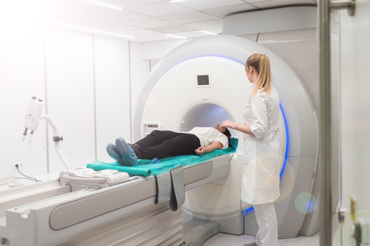
Magnetic resonance imaging (MRI) is universally recognized as the gold standard imaging test for diagnosing herniated discs. This powerful diagnostic tool uses magnetic fields and radio waves to create detailed cross-sectional images of the spine, providing unparalleled visualization of soft tissues including intervertebral discs, the spinal cord, and nerve roots.
The superiority of MRI lies in its ability to distinguish between different types of soft tissue with remarkable clarity. Unlike other imaging modalities, MRI can clearly show the disc’s nucleus pulposus, annulus fibrosus, and any herniated material that may be compressing nearby neural structures. This level of detail is crucial for accurate diagnosis and treatment planning.
Why MRI excels over other imaging methods
Superior soft tissue contrast
MRI’s greatest strength is its exceptional soft tissue contrast resolution. The test can differentiate between healthy disc material, degenerated tissue, and herniated fragments with precision that other imaging methods simply cannot match. This capability allows physicians to assess not only the presence of a herniation but also its size, location, and relationship to surrounding structures.
Multiplanar imaging capabilities
MRI can capture images in multiple planes—sagittal, axial, and coronal—providing a comprehensive three-dimensional understanding of the spine. This multiplanar approach enables physicians to visualize herniations from different angles, ensuring nothing is missed and providing crucial information for surgical planning when necessary.
No radiation exposure
Unlike CT scans and X-rays, MRI uses no ionizing radiation, making it safer for repeated examinations and suitable for patients who require multiple follow-up studies. This safety profile is particularly important for younger patients or those who may need ongoing monitoring of their condition.
Comparing Alternative Imaging Methods
CT scans: the secondary option
Computed tomography (CT) scans serve as a valuable alternative when MRI is contraindicated or unavailable. CT scans excel at visualizing bony structures and can detect disc herniations, particularly when combined with contrast agents (CT myelography). However, CT’s soft tissue resolution is inferior to MRI, and the test involves radiation exposure.
CT scans are often preferred in emergency situations due to their speed and widespread availability. They are also the go-to choice for patients with MRI contraindications, such as certain metallic implants or severe claustrophobia.
X-Rays: limited diagnostic value
Plain radiographs (X-rays) have significant limitations in diagnosing herniated discs. Since discs are composed primarily of soft tissue, they are not directly visible on X-rays. While X-rays can show disc space narrowing, vertebral alignment issues, and degenerative changes, they cannot definitively diagnose a disc herniation.
X-rays are typically used as an initial screening tool to rule out fractures, infections, or other bony abnormalities before proceeding to more advanced imaging.
When Each Imaging Method Is Recommended
First-line MRI indications
MRI is recommended for:
- Persistent back pain with neurological symptoms
- Suspected compression of a nerve root, especially the sciatic nerve
- Planning for surgical intervention
- Monitoring treatment response
- Evaluating complex spinal conditions
CT scan scenarios
CT scans are preferred when:
- MRI is contraindicated due to metallic implants
- Emergency evaluation is needed
- Bony abnormalities need to be assessed
- Patients cannot tolerate MRI due to claustrophobia
- Cost considerations make MRI unavailable
X-ray applications
X-rays remain useful for:
- Initial screening of back pain
- Ruling out fractures or instability
- Assessing overall spinal alignment
- Monitoring hardware in postsurgical patients
Factors Affecting Imaging Test Choice
Patient-specific considerations
Several factors influence the choice of imaging modality. Patient claustrophobia, the presence of metallic implants, kidney function (for contrast studies), and patient physique all play roles in determining the most appropriate test. Pregnant patients require special consideration, with MRI generally preferred over CT due to radiation concerns.
Clinical context
The urgency of diagnosis, availability of equipment, and cost considerations also influence imaging decisions. In acute settings, CT may be chosen for its speed, while outpatient evaluations typically favor MRI for its superior diagnostic accuracy.
Limitations and considerations
Despite its gold standard status, MRI has limitations. The test can be expensive, time-consuming, and unavailable in some settings. Additionally, MRI may reveal incidental findings or asymptomatic disc abnormalities that do not correlate with the patient’s symptoms, potentially leading to overdiagnosis or unnecessary interventions.
The Future of Disc Imaging
Emerging technologies continue to enhance disc imaging capabilities. Advanced MRI sequences, such as diffusion tensor imaging and T2 mapping, provide even more detailed information about disc health and integrity. These developments promise to further solidify MRI’s position as the premier imaging modality for spinal conditions.
Understanding when and why different imaging tests are used empowers patients to have informed discussions with their healthcare providers about the most appropriate diagnostic approach for their specific situation. When a herniated disc is suspected, MRI provides the detailed accurate information necessary for optimal treatment planning and patient outcomes.
If you have a herniated disc that is not responding to conservative treatment, a discectomy or less invasive microdiscectomy may be discussed and potentially recommended. Although this is generally a very successful procedure, having a large hole in the outer ring of the disc more than doubles the risk of needing another operation. A new treatment, Barricaid, is a bone-anchored device that closes this hole, and 95 percent of Barricaid patients did not undergo a reoperation due to reherniation in a 2-year study time frame. This treatment is done immediately following the discectomy—during the same operation—and does not require any additional incisions or time in the hospital.
If you have any questions about the Barricaid treatment, ask your doctor or contact us today.
For full benefit/risk information, please visit: https://www.barricaid.com/instructions.
Comments