

The Straight Leg Raise Test: A Key Diagnostic Tool for Herniated Discs
Herniated discs are a common cause of lower back and leg pain, especially when they compress spinal nerve roots. When patients have lower back pain radiating down their legs, healthcare providers often turn to a time-tested diagnostic tool: the straight leg raise (SLR) test. This basic clinical examination has been a cornerstone of spinal assessment for more than a century, helping doctors identify suspected lumbar disc herniations and nerve root compression.
The SLR test offers clinicians a quick, noninvasive method to assess whether leg pain stems from spinal nerve irritation. While it is straightforward in execution, understanding its proper application and interpretation is crucial for accurate diagnosis and appropriate patient care. In this article, you will learn how the straight leg test works, what a positive or negative result means, and its role in diagnosing herniated discs. You will also gain an understanding of its limitations and how it fits into a comprehensive clinical evaluation.
The Connection between Disc Herniation and Nerve Pain
To appreciate the significance of the SLR test, we must first examine the pathophysiology it helps doctors identify. Lumbar disc herniation occurs when the gel-like center of an intervertebral disc (nucleus pulposus) protrudes through a weakness in the disc’s outer ring (annulus fibrosus). This protrusion can impinge upon adjacent nerve roots, particularly at the L5 and S1 levels.
When nerve roots become compressed or irritated, patients typically experience a constellation of symptoms known as sciatica. This condition manifests as sharp, shooting pain that travels from the lower back down through the buttock and leg, often accompanied by numbness, tingling, or muscle weakness.
Test Methodology and Technique
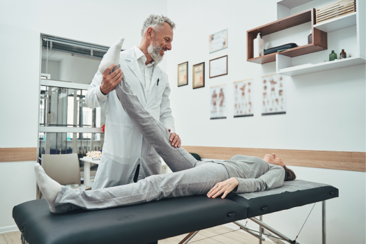
The straight leg raise test was originally described by French neurologist Charles Lasègue in the 1800s. The test works by creating tension on the lumbosacral nerve roots through hip flexion while maintaining knee extension. This movement stretches the sciatic nerve and its nerve roots, particularly affecting the L4, L5, and S1 nerve roots. When a herniated disc compresses these nerve roots, the additional tension created during the test reproduces the patient’s symptoms, indicating possible nerve root irritation or compression.
The examination begins with the patient lying on their back on an examination table. The patient should be as relaxed as possible, with both legs initially resting flat on the table.
The healthcare provider performs the test by following these specific steps:
- Initial assessment - The examiner stands beside the patient and grasps the ankle and heel of the patient’s affected leg.
- Gradual hip flexion - While keeping the patient’s knee completely straight, the examiner slowly raises the leg by flexing the hip. The movement should be smooth and controlled, typically raising the leg at a rate of approximately 1–2 degrees per second.
- Angle monitoring - The examiner continues raising the leg while monitoring the angle of hip flexion, paying particular attention to the critical range between 30 and 70 degrees.
- Symptom assessment - Throughout the procedure, the examiner asks the patient to report any pain, numbness, tingling, or other symptoms, particularly noting when symptoms begin and their location.
- Documentation - The examiner records the angle at which symptoms occur, the nature of the symptoms, and their distribution pattern.
Interpreting Test Results
Positive test findings
A positive straight leg raise test occurs when the patient experiences typical leg pain or neurological symptoms when the affected leg is raised between 30 and 70 degrees. The quality and distribution of reproduced symptoms provide additional diagnostic clues. Sharp, shooting pain that follows the sciatic nerve, radiating from the lower back down the posterior or lateral aspect of the leg and potentially extending to the foot, is more significant than vague discomfort or isolated back pain.
Negative test findings
When leg elevation fails to reproduce radicular symptoms, the test is considered negative. This finding suggests that nerve root compression may not be the primary source of pain, although it does not completely exclude the possibility of disc herniation, as some herniated discs may not cause sufficient nerve root tension to produce positive test results.
Clinical Performance and Diagnostic Value
The SLR test demonstrates strong screening capabilities but has important limitations that clinicians must understand.
Sensitivity characteristics
Research indicates that the SLR test exhibits high sensitivity, ranging from 80 percent to 97 percent for detecting nerve root involvement. This high sensitivity makes it particularly valuable as a screening tool—a negative test effectively reduces the likelihood of significant nerve root compression.
Specificity limitations
The test’s specificity is considerably lower, typically falling between 40 percent and 57 percent. This means positive results may occur in various conditions beyond disc herniation, including:
- Hip joint pathology
- Piriformis syndrome
- Hamstring muscle tightness
- Peripheral nerve disorders
- Spinal tumors
Clinical implications
These performance characteristics position the SLR test as an excellent “rule-out” tool rather than a definitive diagnostic test. Healthcare providers must integrate SLR findings with comprehensive clinical assessment for accurate diagnosis.
Complementary Diagnostic Approaches
Given the SLR test’s limitations, additional evaluation methods are often necessary for complete assessment:
Advanced imaging
Magnetic resonance imaging (MRI) remains the gold standard for visualizing disc structure and assessing the degree of nerve root compression. MRI provides detailed anatomical information that complements clinical findings from the SLR test.
Neurological assessment
Comprehensive neurological examination, including muscle strength testing, reflex evaluation, and sensory mapping, provides crucial additional data points that support or refute SLR findings.
Electrophysiological studies
In cases where results of clinical tests remain unclear or symptoms persist despite treatment, electromyography (EMG) and nerve conduction studies can localize nerve dysfunction and assess severity.
Common Pitfalls and False Results
Understanding potential sources of error helps clinicians interpret SLR results more accurately:
False positive scenarios
As mentioned above in the discussion of specificity, several conditions can mimic positive SLR tests:
- Tight hamstring muscles limiting hip flexion
- Hip joint arthritis or impingement
- Sacroiliac joint dysfunction
- Psychological factors or symptom magnification
False negative possibilities
Certain circumstances may produce negative results despite significant pathology:
- Central disc herniations without lateral nerve root compression
- High pain tolerance that masks symptom reproduction
- Medications that suppress pain perception
- Chronic adaptations that reduce acute symptom response
Clinical Applications and Best Practices
The SLR test is widely used in a variety of healthcare settings, from primary care offices to specialized spine clinics. Its simplicity and minimal equipment requirements make it accessible to practitioners at all levels to determine appropriate treatment strategies:
- Conservative management - Negative or mildly positive tests may indicate suitability for nonsurgical treatment approaches
- Surgical consultation - Strongly positive tests with severe neurological symptoms may warrant surgical evaluation
- Monitoring progress - Serial testing can assess improvement or deterioration over time
Effective use of the SLR test requires integration with patient history, symptom patterns, and physical examination findings. Isolated reliance on test results without considering the broader clinical picture can lead to diagnostic errors.
When discussing SLR results with patients, providers should explain both the test’s value and its limitations. Patients benefit from understanding additional testing may be necessary regardless of initial results.
Maximizing Diagnostic Value
The straight leg raise test serves as a valuable tool in diagnosing suspected lumbar disc herniation. Its high sensitivity makes it particularly useful for screening patients and guiding further evaluation decisions. However, clinicians must remember no single test provides definitive answers in complex spinal conditions.
Healthcare providers must understand both the capabilities and limitations of this test, interpreting results within the broader clinical context rather than relying on it as a stand-alone diagnostic tool. When performed correctly and interpreted appropriately, the straight leg raise test continues to serve as a cornerstone of spinal examination, helping millions of patients receive appropriate care for their lower back and leg pain symptoms.
If you have a herniated disc that is not responding to conservative treatment, a discectomy or less invasive microdiscectomy may be discussed and potentially recommended. Although this is generally a very successful procedure, having a large hole in the outer ring of the disc more than doubles the risk of needing another operation. A new treatment, Barricaid, is a bone-anchored device that closes this hole, and 95 percent of Barricaid patients did not undergo a reoperation due to reherniation in a 2-year study time frame. This treatment is done immediately following the discectomy—during the same operation—and does not require any additional incisions or time in the hospital.
If you have any questions about the Barricaid treatment, ask your doctor or contact us today.
For full benefit/risk information, please visit: https://www.barricaid.com/instructions.
Comments