

Understanding Disc Herniation: Types and Classifications
Herniated discs represent one of the most common causes of back pain and neurological symptoms affecting millions of adults worldwide. While the general public may think all disc herniations are the same, they are actually categorized into various types. These distinctions matter, as they influence symptom assessment, prognosis, and treatment strategies. Understanding the various types and classifications of herniated discs is crucial for both patients and healthcare providers to determine appropriate treatment strategies and predict outcomes.
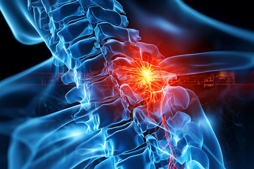
A herniated disc occurs when part or all of the nucleus pulposus (the gel-like center of the disc) protrudes through a tear or weakness in the annulus fibrosus (the tough outer ring of the disc). This can compress the nerves or spinal cord, causing pain and spinal cord dysfunction. The classification of herniated discs has evolved significantly over the years, with multiple systems developed to categorize these injuries based on various factors, including anatomical location and degree of disc displacement. In this article, we dive deeper into the major types of herniated discs, how they are classified, and what this means for individuals living with them.
Types Classified by Anatomical Location
Cervical spine herniation
In the cervical spine, the C6-7 is the most common herniated disc that causes symptoms, particularly radiculopathy. Patients experience neck pain that may radiate into the shoulder, arm, and hand, often accompanied by specific patterns of weakness and sensory changes, depending on which nerve root is compressed.
Thoracic spine herniation
Thoracic disc herniations are less common than cervical or lumbar herniations due to the biomechanical stability provided by the rib cage. These herniations are often accompanied by atypical symptoms and may be discovered incidentally on imaging studies.
Lumbar spine herniation
Lumbar disc herniations are the most frequent type, with approximately 95 percent of herniated discs occurring in patients between 25 and 55 years old. This type of herniation is the most common due to the mechanical stresses placed on the lower back.
Morphological Classification System
Another widely accepted classification system categorizes herniated discs based on their morphological appearance and the extent of nuclear material displacement. This system includes four primary stages: disc bulge, disc protrusion, disc extrusion, and disc sequestration. The first two stages are referred to as incomplete, while the last two are considered complete herniations.
Disc bulge
A disc bulge represents the mildest form of disc pathology, where the annulus fibrosus remains intact but the disc extends beyond its normal boundaries. This is often considered a normal part of the aging process rather than a true herniation and may not cause symptoms in many patients.
Disc protrusion
In a protrusion, the nucleus pulposus (the gel-like center) pushes against the annulus fibrosus (the tough outer ring), causing it to bulge outward, but the outer fibers of the annulus remain intact.
Disc extrusion
Extrusion occurs when the nucleus pulposus breaks through the annulus fibrosus but remains connected to the disc space. Extrusions represent a more severe form of herniation than protrusions and are more likely to cause significant symptoms.
Disc sequestration
Sequestration represents the most severe form of disc herniation. Disc sequestration is an extrusion where the disc material migrates and becomes separated from the rest of the herniation. In this type, a fragment of the nucleus pulposus breaks free entirely and may migrate within the spinal canal, potentially causing severe nerve compression.
Diagnosing Herniated Discs
Imaging tests
MRI is the preferred and most sensitive test for visualizing herniated discs. However, it is important to note that imaging findings do not always correlate with symptoms. Many individuals with disc herniations that are visible on MRI studies have no symptoms at all.
Clinical assessment
Clinical evaluation remains the primary way to determine the significance of disc herniations. The straight leg raise test and the contralateral (crossed) straight leg raise test are important clinical tools for assessing lumbar disc herniations, with the crossed straight leg raise test having a specificity higher than 90 percent.
Clinical Significance and Treatment Implications
The type and classification of herniated discs significantly influence treatment decisions and prognosis. Research has shown that different types of herniations have varying rates of spontaneous regression. In one systematic review, the rate of spontaneous regression was found to be 96 percent for disc sequestration, 70 percent for disc extrusion, 41 percent for disc protrusion, and 13 percent for disc bulging.
This finding is particularly important because it suggests that more severe herniations (sequestrations) are actually more likely to resolve naturally without surgical intervention, likely due to increased inflammatory response and subsequent resorption of the herniated material.
Conservative Treatment
In more than 85 percent of patients, symptoms associated with an acute herniated disc will resolve within 8 to 12 weeks without any specific treatments. Therefore, the initial approach is conservative and typically includes:
- Non-steroidal anti-inflammatory drugs (NSAIDs)
- Physical therapy (after initial acute phase)
- Activity modification
- Epidural corticosteroid injections
Surgical Considerations
Surgery is typically reserved for patients who:
- Do not benefit from conservative treatment after 6–12 weeks
- Show symptoms of progressive neurological deficits
- Develop cauda equina syndrome (emergency situation)
- Experience severe, intractable pain affecting quality of life
Future Directions and Emerging Classifications
As our understanding of disc pathology continues to evolve, new classification systems are being developed that incorporate advanced imaging techniques, genetic factors, and inflammatory markers. These emerging systems may provide better prediction of treatment outcomes and personalize therapy approaches.
The integration of artificial intelligence and machine learning in radiology is also contributing to more objective and reproducible classification systems, potentially reducing inter-observer variability in disc herniation assessment.
The key takeaway for both patients and healthcare providers is that disc herniation classification is a practical tool that directly influences treatment decisions. The encouraging finding that more severe herniations often have better spontaneous resolution rates underscores the importance of accurate classification and appropriate conservative management in many cases.
As research continues to refine our understanding of disc pathology, these classification systems will likely continue to evolve, ultimately leading to more personalized and effective treatment strategies for patients suffering from this common and often debilitating condition.
If you have a herniated disc that is not responding to conservative treatment, a discectomy may be discussed and potentially recommended. Although this is generally a very successful back surgery procedure, having a large hole in the outer ring of the disc more than doubles the risk of needing another operation. A new treatment, Barricaid, is a bone-anchored device that closes this hole, and 95 percent of Barricaid patients did not undergo a reoperation due to reherniation in a 2-year study timeframe. This treatment is done immediately following the discectomy—during the same operation—and does not require any additional incisions or time in the hospital.
If you have any questions about the Barricaid treatment, ask your doctor or contact us directly.
For full benefit/risk information, please visit: https://www.barricaid.com/instructions.
Comments