

Depending on who you ask, an MRI can either be necessary or not beneficial when it comes to diagnosing herniated spinal discs. This article takes a closer look at times when an MRI may be beneficial and when it is not something that is necessary.
MRIs May Not Be Beneficial with Sciatica
According to research reported by the Journal of Neurosurgery: Spine, MRI scans were not helpful with treatment-related decisions for sciatica, which is often related to a herniated disc. Specifically, an MRI did not make much of a difference in determining when to consider surgery and when to continue with conservative treatments such as physical therapy.
MRIs Can Be Beneficial if the Source of Discomfort Is Unclear
There are many issues with the spine that can contribute to herniated disc symptoms. If there are doubts about whether there may be other sources of discomfort, your doctor may order an MRI scan. An MRI is an effective way to determine if there are structural or soft tissue problems, and it may also be necessary if you will be having surgery for your herniated disc. MRIs are often done to ensure the correct location is treated.
Accurate Diagnosis Is Possible without an MRI
Herniated spinal discs have classic symptoms that tend to be consistently reliable when it comes to making a diagnosis, so you need to be descriptive about your symptoms during your initial appointment with your doctor or a spine specialist. If the doctor suspects you have a herniated disc, he or she may advise you to begin nonsurgical treatments to see if your symptoms improve over the next few weeks. When a doctor diagnoses a herniated disc without an MRI, the diagnosis is usually based on the following factors:
• The nature of your symptoms
• When your pain is more intense
• The triggers for your discomfort
• Whether you have lower back pain that extends down into your legs
• The causes of improving your symptoms and what makes them worse
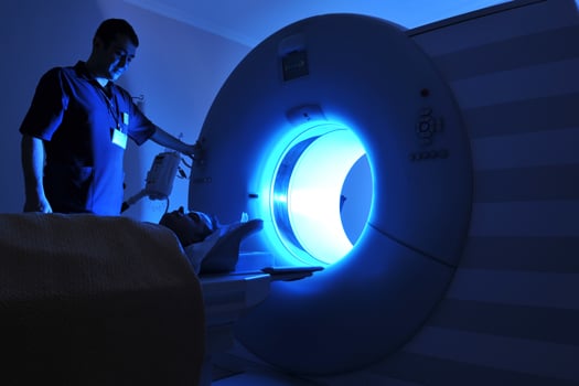
An MRI Provides Detailed Images that Can Aid Diagnosis
An MRI that is ordered to accurately diagnose a herniated disc is a process that provides detailed results. When the scan is performed, you will rest on a bed that is eased into the scanner, which is operated by a radiographer. Radiographers are trained to ensure the correct images are taken so they can be reviewed by a physician or spine specialist.
An MRI goes beyond what is typically seen with an X-ray, which does not show soft tissues. However, an MRI scan is capable of showing detailed images of soft tissues. This allows possible sources of your discomfort to be identified with a fair degree of certainty.
If you have a herniated disc that is not responding to conservative treatment, a discectomy may be discussed and potentially recommended. Although this is generally a very successful procedure, having a large hole in the outer ring of the disc more than doubles the risk of needing another operation. A new treatment, Barricaid, is a bone-anchored device that closes this hole, and 95 percent of Barricaid patients did not undergo a reoperation due to reherniation in a 2-year study time frame. This treatment is done immediately following the discectomy—during the same operation—and does not require any additional incisions or time in the hospital.
If you have any questions about the Barricaid treatment, ask your doctor or contact us at 844-705-1081.
For full benefit/risk information, please visit: https://www.barricaid.com/instructions.
Comments