

Hemilaminectomy and Microdiscectomy: Your Guide to Combined Spine Surgery
A hemilaminectomy with microdiscectomy is a sophisticated spinal surgery that combines two complementary procedures to address multiple spinal conditions simultaneously. This minimally invasive approach has revolutionized the treatment of patients suffering from both spinal stenosis and herniated discs, offering relief from debilitating back pain and neurological symptoms. In this article, we dive deeper into what microdiscectomy with hemilaminectomy is designed to treat, how it works, and what to expect during and after the procedure.
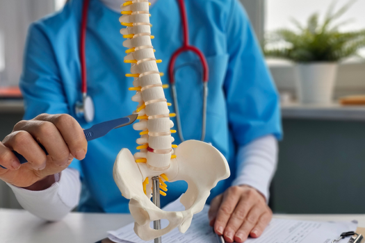
The Basics of Hemilaminectomy with Microdiscectomy
The procedure involves removing a portion of the lamina (the bony arch covering the spinal canal) on one side of the vertebra, followed by the precise removal of herniated disc material that may be compressing nearby nerve roots. This dual approach allows surgeons to address both structural narrowing of the spinal canal and disc-related nerve compression in a single operation.
Hemilaminectomy Explained
A hemilaminectomy, also known as a unilateral laminotomy, involves the surgical removal of part of the lamina on one side of a vertebra. The lamina is the posterior portion of the vertebral arch that protects the spinal cord and nerve roots. When this structure becomes thickened or enlarged due to arthritis, bone spurs, or other degenerative changes, it can narrow the spinal canal and compress neural structures.
During a hemilaminectomy, the surgeon carefully removes only the necessary amount of bone and ligament tissue to decompress the affected nerve roots while preserving spinal stability. This selective approach maintains the structural integrity of the spine while creating adequate space for the compressed nerves to function properly.
Understanding Microdiscectomy
Microdiscectomy is a minimally invasive surgical technique used to remove herniated or protruding disc material that compresses spinal nerves. This type of herniated disc surgery utilizes a surgical microscope and specialized instruments to access the affected disc through a small incision, typically measuring only one to two inches in length.
The surgeon removes only the problematic portion of the disc that is causing nerve compression, leaving healthy disc tissue intact. This precision-focused approach minimizes damage to surrounding structures and promotes faster healing compared to traditional open disc surgery.
Why These Procedures Are Combined
Combining hemilaminectomy with microdiscectomy addresses multiple sources of nerve compression that often occur simultaneously. Many patients with herniated discs also develop spinal stenosis, a condition where the spinal canal narrows due to bone and ligament overgrowth. By performing both procedures during a single surgery, the surgeon can comprehensively treat all sources of neural compression while minimizing the patient’s exposure to surgical risks and reducing recovery time.
This combined approach is particularly beneficial for patients with complex spinal conditions where both bony and soft tissue structures contribute to nerve compression. The hemilaminectomy creates additional space within the spinal canal, while the microdiscectomy removes the specific disc material causing direct nerve root irritation.
Conditions Treated by This Combined Procedure
Hemilaminectomy with microdiscectomy effectively treats several spinal conditions, including lumbar spinal stenosis with concurrent disc herniation, degenerative disc disease with associated spinal narrowing, and spondylolisthesis with disc involvement. Patients typically experience symptoms such as leg pain, numbness, weakness, and difficulty walking, particularly when standing upright or walking for extended periods.
The procedure is most commonly performed in the lumbar spine, where the combination of disc herniation and spinal stenosis frequently occurs. However, it can also be adapted for cervical spine conditions when appropriate.
The Surgical Procedure
The surgery is performed under general anesthesia with the patient positioned face down on the operating table. The surgeon makes a small incision over the affected vertebral level and carefully dissects through the muscle layers to reach the spine. Using a surgical microscope for magnification, the surgeon first performs the hemilaminectomy by removing the necessary bone and ligament tissue to decompress the spinal canal.
Following the decompression, the surgeon proceeds with the microdiscectomy, carefully removing the herniated disc material that compresses the nerve root. Throughout the procedure, the surgeon takes great care to protect the nerve roots and surrounding structures from injury.
Recovery and Rehabilitation
Recovery from hemilaminectomy with microdiscectomy typically involves a structured rehabilitation program designed to restore strength, flexibility, and function. Most patients can expect to return to light activities within a few weeks, with full recovery occurring over several months.
Physical therapy plays a crucial role in the recovery process, helping patients regain strength and mobility while preventing future complications. The rehabilitation program typically begins with gentle range of motion exercises and gradually progresses to more challenging strengthening activities.
Expected Outcomes and Benefits
The combined procedure offers excellent outcomes for appropriately selected patients, with most experiencing significant improvement in pain and neurological symptoms. The minimally invasive nature of the surgery results in less tissue damage, reduced postoperative pain, and faster recovery compared to traditional open procedures.
Studies demonstrate high success rates for this combined approach, with most patients achieving substantial pain relief and functional improvement. The preservation of spinal stability through the selective removal of tissue contributes to long-term positive outcomes.
Hemilaminectomy with microdiscectomy represents a sophisticated solution for patients suffering from complex spinal conditions involving both bony and disc-related nerve compression. This combined minimally invasive approach offers the advantage of addressing multiple sources of symptoms in a single procedure while minimizing surgical risks and recovery time. For patients with appropriate conditions, this procedure can provide significant relief from debilitating symptoms and restore quality of life.
Although discectomy surgery is generally a very successful procedure, a hole is left in the outer wall of the disc. Patients with a large hole in the outer ring of the disc are more than twice as likely to reherniate after surgery. Barricaid is a bone-anchored device designed to reduce reherniations by closing the large hole often left in the spinal disc after microdiscectomy. In a large-scale study, 95 percent of Barricaid patients did not undergo a reoperation due to reherniation in the 2-year study timeframe. This treatment is performed immediately following the discectomy—during the same operation—and does not require any additional incisions or time in the hospital.
If you have any questions about the Barricaid treatment or how to get access to Barricaid, ask your doctor or contact us at 844-288-7474.
For full benefit/risk information, please visit: https://www.barricaid.com/instructions.
Comments