

Microdiscectomy is a minimally invasive surgical procedure that relieves sciatic nerve pain by removing the portion of a herniated disc compressing a spinal nerve root. It resolves the physical cause of sciatica, not just the symptoms. In this article, we take a closer look at the science behind how this procedure works, who benefits most, and what recovery looks like.
What Is the Root Cause of Sciatic Nerve Pain?
Sciatic nerve pain originates when a herniated intervertebral disc pushes against one of the nerve roots that form the sciatic nerve, most commonly at the L4–L5 or L5–S1 spinal levels. The disc (a soft, gel-filled cushion between vertebrae) can rupture under sustained mechanical pressure, age-related degeneration, or acute injury. When the nucleus pulposus (the inner gel material) escapes the disc’s outer casing, it intrudes into the spinal canal and presses directly against nerve tissue.
That pressure produces the hallmark symptoms of sciatica: sharp, radiating pain down one leg, numbness, tingling, and, in severe cases, muscle weakness. The nerve compression also triggers a localized inflammatory response. The immune system identifies the nucleus pulposus as a foreign substance and releases pro-inflammatory cytokines, which compound the pain signal. Microdiscectomy addresses both problems simultaneously: it eliminates the mechanical compression and removes the tissue driving inflammation.
How Does Microdiscectomy Physically Remove Pressure from a Compressed Nerve?
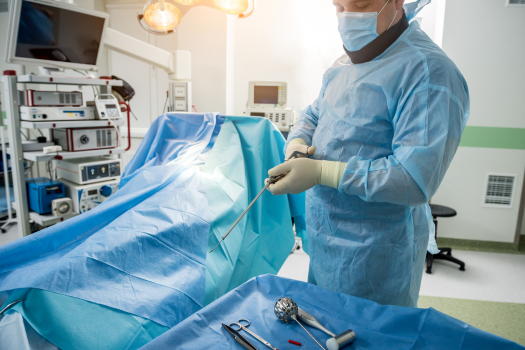
Microdiscectomy removes the herniated disc fragment pressing against the nerve root through a small incision, typically less than one inch, in the lower back. The surgeon uses a surgical microscope or magnifying loupes to visualize the operative field with precision, displacing, not cutting, the paraspinal muscles to reach the spine.
Once the lamina (the bony arch of the vertebra) is exposed, the surgeon removes a small portion of it in a step called a laminotomy. This creates a window into the spinal canal. The nerve root is then carefully retracted to one side, and the offending disc material is extracted using specialized grasping instruments called rongeurs. The procedure targets only the herniated fragment, leaving the majority of the disc intact to preserve spinal stability. Because the muscles are spread rather than cut, structural damage to the surrounding tissue is minimal.
Why Does Minimally Invasive Technique Matter for Nerve Recovery?
Minimally invasive technique matters because it dramatically reduces trauma to the soft tissue, bones, and ligaments that support the spine. Traditional open discectomy requires cutting through layers of muscle to access the spine, which increases blood loss, prolongs recovery, and elevates the risk of postoperative instability. Microdiscectomy, by contrast, preserves the integrity of those structures.
From a neurological standpoint, the sooner mechanical compression is relieved, the better the nerve’s recovery trajectory. Prolonged compression causes demyelination (breakdown of the protective sheath around nerve fibers) and, in severe cases, axonal damage that is not fully reversible. The precision of microdiscectomy allows surgeons to decompress the nerve quickly and accurately, giving it the best possible environment to heal. Reduced surrounding tissue injury also means less postoperative inflammation near the nerve itself, which further supports recovery.
What Happens to Sciatic Nerve Function after the Herniated Material Is Removed?
After disc material is removed, nerve function typically begins to return within days to weeks, though the timeline depends on how long the nerve was compressed and the degree of injury sustained. The nerve root, now free from mechanical pressure, begins to recover its normal signal transmission.
For most patients, pain relief is the first and most dramatic improvement, often apparent within the first 24 to 48 hours after surgery. Numbness and tingling generally resolve over subsequent weeks. Muscle weakness, when present before surgery, takes the longest to improve because motor nerve fibers require more time and resources to regenerate. In cases where compression was prolonged, some residual sensory changes may persist, though they typically diminish over several months as the nerve undergoes remyelination and axonal repair.
Who Is the Ideal Candidate for Microdiscectomy, and Who Is Not?
The ideal candidate for microdiscectomy is a patient with a confirmed single-level lumbar disc herniation causing persistent radicular leg pain (sciatica) that has not responded to six or more weeks of conservative treatment, including physical therapy, anti-inflammatory medications, and targeted injections. Imaging (typically an MRI) must confirm the disc herniation correlates directly with the patient’s symptoms.
Patients in the last stages of sciatica or with red-flag symptoms, such as progressive neurological deficits or signs of cauda equina syndrome, may require more urgent surgical evaluation rather than completing a course of conservative treatment. Microdiscectomy is generally not the appropriate procedure for patients whose primary complaint is axial low back pain without significant leg symptoms, for those with spinal instability, or for those with multilevel disc disease requiring more extensive reconstruction. Age, overall health, and bone density are also evaluated to ensure the patient can tolerate anesthesia and recover safely.
What Does the Scientific Evidence Say about Long-Term Outcomes?
The scientific evidence for microdiscectomy is robust. Multiple randomized controlled trials and long-term outcome studies consistently show microdiscectomy produces faster and more complete relief of leg pain than continued conservative management for patients with confirmed disc herniation and persistent sciatica. The landmark SPORT (Spine Patient Outcomes Research Trial), published in JAMA in 2006, enrolled 743 patients across 13 multidisciplinary spine clinics and found that surgically treated patients achieved significantly greater improvements in pain, physical function, and disability scores at two-year follow-up compared to those who received only nonoperative care. A subsequent four-year analysis from the same trial confirmed surgical patients maintained greater improvement across all primary and secondary outcomes.
Recurrence rates (meaning reherniation at the same disc level) range from approximately five to twenty-one percent following primary discectomy, with reherniation representing the leading cause of reoperation, according to a large-scale peer-reviewed study drawing on national patient data. Reoperation rates remain low when patients are appropriately selected and adhere to postoperative rehabilitation. Long-term studies (including the Maine Lumbar Spine Study) tracking patients at ten years or more confirm the majority maintain durable relief and do not require additional spinal surgery at the treated level.
Frequently Asked Questions
How long does a microdiscectomy procedure typically take to complete?
Most microdiscectomy procedures take between 45 minutes and two hours, depending on the complexity of the herniation and the patient’s anatomy.
Is microdiscectomy performed under general or local anesthesia?
Usually, general anesthesia is used, though some surgeons perform the procedure under spinal anesthesia for patients who are poor candidates for general anesthesia.
How soon after microdiscectomy can patients return to normal activity?
Most patients return to light activity within a few weeks and resume full physical function, including non-strenuous work, within several weeks. Physically demanding jobs or activities may require several weeks to months.
Does microdiscectomy address the underlying cause of disc herniation or only the symptoms?
It removes the herniated fragment causing compression, which resolves the immediate cause of nerve pain. It does not reverse disc degeneration or eliminate the risk of future herniations at other levels.
What is the difference between microdiscectomy and a standard open discectomy?
Microdiscectomy uses a smaller incision, a surgical microscope, and muscle-sparing technique, resulting in less tissue trauma, shorter hospital stays, and faster recovery than traditional open discectomy.
If you have had a discectomy or microdiscectomy to relieve sciatica caused by a herniated disc, you may experience recurrent sciatica if the disc becomes reherniated, which often occurs if there is a large hole in the outer ring of the disc after surgery. Fortunately, there is a new treatment available to help avoid this. Barricaid is a bone-anchored annular closure device designed to reduce the likelihood of reherniation by closing the hole in the disc after a discectomy, and 95 percent of Barricaid patients did not undergo a reoperation due to reherniation in a 2-year study timeframe. This treatment is done immediately following the discectomy—during the same operation—and does not require any additional incisions or time in the hospital.
To learn more about the Barricaid treatment, ask your doctor or contact us.
For full benefit/risk information, please visit: https://www.barricaid.com/instructions.
Comments