

Exploring the Anatomy of a Discectomy: What Happens to Your Herniated Disc?
A discectomy is a surgical procedure in which a surgeon removes the damaged protruding portion of a spinal disc that is pressing on a nearby nerve. The disc itself is not typically removed in full. Rather, only the herniated nucleus material is extracted, relieving nerve compression and restoring function. In this article, we explore precisely what happens to your herniated disc during a discectomy, from the moment surgery begins to the biological changes that follow.
What Is a Herniated Disc and Why Does It Require Surgery?
A herniated disc occurs when the soft gel-like nucleus pulposus at the center of a spinal disc pushes through a tear in the harder outer ring, called the annulus fibrosus. This displaced material can press directly against a spinal nerve root or the spinal cord itself, causing pain, numbness, or weakness that radiates into the arms or legs depending on the location of the herniation.
Most herniated discs resolve with conservative treatments such as physical therapy, anti-inflammatory medications, and targeted injections. Surgery becomes necessary when nerve compression is severe, when neurological symptoms worsen, or when pain persists for an extended period without meaningful improvement despite conservative treatment. A discectomy or microdiscectomy is generally the preferred surgical option because it is minimally invasive and precisely targeted.
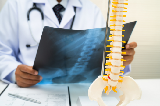
How Do Surgeons Access the Herniated Disc without Damaging the Spine?
Surgeons access the herniated disc through a small incision in the back, typically one to two inches in length, positioned directly over the affected spinal level. A series of dilating tubes are inserted through the muscle tissue rather than cutting through it, which preserves the integrity of surrounding musculature and reduces recovery time significantly.
Using a surgical microscope or endoscope for magnification, the surgeon navigates to the lamina, the bony arch that covers the back of the spinal canal. A small window is then created in the lamina through a technique called a laminotomy, providing a clear line of sight to the compressed nerve root and the bulging disc material beneath it. Fluoroscopic X-ray imaging is used intraoperatively to confirm the correct spinal level before any tissue is removed.
What Portion of the Disc Does a Surgeon Remove during a Discectomy?
The surgeon removes only the herniated fragment of the nucleus pulposus that is directly compressing the nerve. This is a targeted extraction, not a full disc removal. Using specialized instruments such as Kerrison rongeurs and pituitary forceps, the surgeon carefully excises the displaced tissue while the nerve root is gently retracted to one side.
In some cases, the surgeon will also remove loose or degenerated disc material from inside the disc space itself to reduce the likelihood of reherniation. However, the annulus fibrosus and the healthy portions of the disc are left intact whenever possible. The goal is always to decompress the nerve with the least disruption to the surrounding anatomy.
Does the Disc Heal or Regenerate after Herniated Material Is Removed?
The disc does not regenerate in the same way soft tissue elsewhere in the body does. Intervertebral discs have a very limited blood supply, which restricts their capacity for repair. After a discectomy, the disc space generally remains slightly reduced in height compared to a healthy disc, and the annulus fibrosus tear that allowed the herniation to occur may not fully seal.
That said, research published in the European Spine Journal has documented that the remaining disc tissue stabilizes over time and that patients typically experience substantial pain relief and functional improvement following surgery. The body adapts by depositing fibrous scar tissue around the operative site, which provides some structural support. Patients are encouraged to strengthen the surrounding musculature through rehabilitation to compensate for any residual disc vulnerability.
What Are the Structural Differences between a Microdiscectomy and a Standard Discectomy?
A microdiscectomy and a standard open discectomy achieve the same anatomical outcome: removal of herniated disc material to decompress a spinal nerve. The primary distinction lies in how the surgeon reaches the disc. A standard discectomy involves a larger incision and more retraction of the paraspinal muscles, while a microdiscectomy uses a smaller incision, tubular retractors, and microscopic visualization.
Because muscle damage is minimized in microdiscectomy, the procedure is associated with shorter hospital stays, less postoperative pain, and faster return to normal activity. According to a study published in Neurosurgery, outcomes between the two techniques at one year are generally comparable in terms of leg pain relief and neurological recovery, but early recovery metrics favor the minimally invasive approach.
What Happens to the Nerve Root Once Disc Pressure Is Relieved during Surgery?
Once the compressing disc fragment is removed, the nerve root is immediately decompressed and is visually confirmed to move freely in the operative field. In many cases, the surgeon can observe the nerve root physically relax and shift position once pressure is no longer applied.
Nerve recovery after decompression depends on several factors, including how long the nerve was compressed and the severity of the compression prior to surgery. Nerves that were compressed for shorter durations generally recover more quickly and completely. Patients who undergo discectomy within a few months of symptom onset typically experience faster resolution of numbness and weakness than those who delay surgery significantly. Full neurological recovery can take weeks to several months as the nerve regenerates its protective myelin sheath.
What Physical Changes in the Spine Are Visible on Imaging after a Discectomy?
Postoperative MRI scans typically show resolution of the herniation at the treated level, with the nerve root no longer displaced or compressed. The disc space may appear slightly reduced in height compared to preoperative imaging, which is a normal finding and does not indicate a complication.
Scar tissue formation, known as epidural fibrosis, is commonly visible on imaging in the weeks and months following surgery. While this is a standard part of healing, in rare cases, extensive scar tissue can adhere to the nerve root and contribute to residual or recurring symptoms. Surgeons generally take care to minimize tissue trauma during the procedure to reduce the likelihood of problematic fibrosis.
A discectomy is a precise and well-established surgical intervention designed to address the specific anatomical problem of a herniated disc compressing a spinal nerve. By removing only the displaced disc material responsible for the compression, surgeons restore nerve function with minimal disruption to the surrounding structures. Understanding the anatomy of the procedure, from the method of access to the biological response of the disc and nerve, helps patients approach surgery with realistic expectations and a clearer sense of what discectomy recovery involves.
If you are considering a discectomy, a board-certified spine surgeon can evaluate your imaging, review your symptom history, and determine whether surgical intervention is appropriate for your specific anatomy and clinical presentation.
Frequently Asked Questions
Is the entire disc removed during a discectomy?
No. Only the herniated portion of the nucleus pulposus that is pressing on the nerve is removed. The remainder of the disc is preserved.
How long does it take for the nerve to recover after a discectomy?
Often, pain relief begins within days, but full neurological recovery of numbness or weakness can take weeks to several months depending on how long the nerve was compressed.
Can a herniated disc return after a discectomy?
Yes. Reherniation at the same disc level occurs in approximately five to fifteen percent of patients, most commonly within the first few months after surgery.
Is a discectomy performed under general anesthesia?
Generally, yes. Most discectomies are performed under general anesthesia, though spinal or regional anesthesia is used in some clinical settings depending on patient health factors.
How soon after a discectomy do most patients return to work?
Most patients with desk-based jobs return to work within two to four weeks; those in physically demanding roles typically require six to twelve weeks of recovery before resuming full duties.
Even though discectomy surgery is a common and generally quite successful form of herniated disc surgery, a hole is frequently left in the outer wall of the disc. In fact, patients with a large annular defect are more than twice as likely to experience a reherniation. These reherniations often require additional surgery or even fusions. Fortunately, there is a treatment specifically designed to close the large holes that are often left in spinal discs after discectomy surgery. Barricaid is a bone-anchored device designed to reduce the likelihood of reherniation by closing the large hole often left in the spinal disc after discectomy, and 95 percent of Barricaid patients did not undergo a reoperation due to reherniation in a 2-year study timeframe. This treatment is done immediately following the discectomy—during the same operation—and does not require any additional incisions or time in the hospital.
If you have any questions about the Barricaid treatment or how to get access to Barricaid, ask your doctor or contact us today.
For full benefit/risk information, please visit: https://www.barricaid.com/instructions.
Comments