

Microdiscectomy and Bone Removal: What You Need to Know
Microdiscectomy is a minimally invasive surgery that removes part or all of a herniated disc that is pressing on a nerve and causing pain, numbness, tingling, or weakness in the back, neck, arms, or legs. Microdiscectomy can relieve the symptoms of sciatica, spinal stenosis, and other spinal conditions and improve the function and mobility of the spine.
But how much bone is removed during microdiscectomy? Is it a significant amount that can affect the stability or integrity of the spine? In this article you will answers to these questions and more by providing a detailed explanation of the procedure and the amount of bone that is removed during microdiscectomy.
Understanding Microdiscectomy
Microdiscectomy is a type of discectomy, which is surgery that removes part or all of a spinal disc that is herniated or damaged. A spinal disc is a soft, cushion-like structure that sits between the vertebrae, the bones that make up the spine. Each spinal disc has two layers: a tough outer layer called the annulus fibrosus and a gel-like inner layer called the nucleus pulposus. Disc herniation occurs when the nucleus pulposus leaks out through a tear or a crack in the annulus fibrosus and compresses or irritates a nearby nerve root or the spinal cord.
A microdiscectomy is different from a traditional open discectomy, which involves a larger incision and more muscle and tissue disruption. A microdiscectomy involves making a smaller incision—about 1 to 1.5 inches long—and using a special microscope or magnifying device to visualize the affected area. The surgeon also uses special instruments and techniques to minimize the damage to the surrounding structures and preserve as much of the healthy disc as possible.
The Procedure
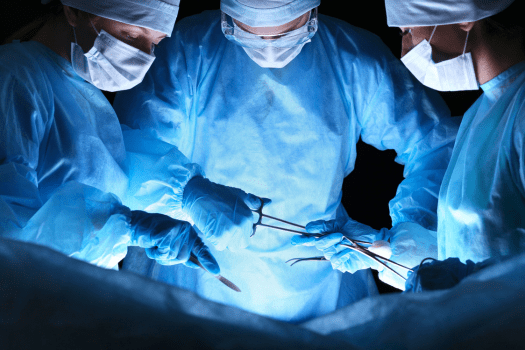
A microdiscectomy is performed under general anesthesia, which means the patient is asleep and does not feel any pain during the surgery. The patient lies face down on a special table that allows the surgeon to adjust the position of the spine. The surgeon makes a small incision in the middle of the back, over the affected disc, and then moves the back muscles aside, without cutting them, to expose the spine. The surgeon may use an X-ray or a fluoroscope, a device that projects live images of the spine on a screen, to locate the exact level of the disc herniation.
The surgeon may remove a small portion of the bone that covers the nerve root or the spinal cord. This bone is called the lamina, and the procedure is called a laminectomy. The surgeon may also remove a small part of the facet joint, the bony projection that connects the vertebrae, to create more space for the nerve.
The surgeon uses a special hook or retractor to gently pull the nerve root away from the disc and then uses small forceps or a suction device to remove the herniated disc material that is compressing the nerve. Then the surgeon may also use a laser or a radiofrequency device to shrink or vaporize any remaining disc material or scar tissue. The surgeon then releases the nerve root, checks for any bleeding or leakage of spinal fluid and closes the incision with stitches or staples
Bone Removal in Microdiscectomy
The amount of bone removed during microdiscectomy is usually very small and does not affect the stability or integrity of the spine. The bone that is removed is only a thin layer of the lamina and sometimes a small part of the facet joint. The amount of bone removed depends on the size and location of the disc herniation and the anatomy of the patient. The surgeon only removes as much bone as necessary to access and decompress the nerve and prevent any recurrence of disc herniation.
The removal of a small amount of bone during microdiscectomy does not require any bone grafting or fusion, which are procedures that involve placing bone or metal implants to join or stabilize the vertebrae. Bone grafting or fusion may be needed in cases of severe disc degeneration, instability, or deformity, which are not common indications for microdiscectomy. The removal of a small amount of bone during microdiscectomy also does not cause any significant loss of height or mobility of the spine, as the disc and the vertebrae remain largely intact.
Postoperative Recovery
Minimizing bone removal in microdiscectomy has positive implications for postoperative recovery. Patients often experience less pain, reduced risk of complications, and a faster return to normal activities. The preservation of spinal stability also contributes to long-term wellbeing, minimizing the likelihood of future issues.
Individualized Approach
Microdiscectomy is not a one-size-fits-all procedure. Surgeons carefully evaluate each patient’s condition and tailor the intervention to address specific needs. This individualized approach ensures bone removal is kept to a minimum while effectively treating the herniated disc.
The amount of bone removed during microdiscectomy is a crucial aspect of the surgical process. By understanding the purpose, procedure, and implications of bone removal, patients can make informed decisions about their treatment.
Advances in surgical techniques continue to refine the precision of microdiscectomy, emphasizing the importance of preserving spinal stability while addressing the root cause of pain. If you are considering microdiscectomy surgery, consult with your healthcare provider to discuss the specific details of your case and the anticipated bone removal involved in your surgery.
Whether a patient has a traditional discectomy, a less invasive microdiscectomy, or spinal fusion surgery, recovery time, pain levels, and other post-surgery issues vary among individuals. An additional factor affecting outcomes is a large hole in the outer ring of the disc after surgery. If the hole in the disc is larger than a standard pencil eraser, the patient has a significant risk of experiencing a reherniation. Patients with a large hole in the outer ring of the disc are more than twice as likely to reherniate after surgery. These reherniations often require additional surgery or even a larger spinal fusion operation. Barricaid is a bone-anchored device shown to reduce reherniations by closing the hole in the disc after a discectomy, and 95 percent of Barricaid patients did not undergo a reoperation due to reherniation in a 2-year study timeframe. This treatment is done immediately following the discectomy—during the same operation—and does not require any additional incisions or time in the hospital.
If you have any questions about the Barricaid treatment or how to get access to Barricaid, you may ask your doctor or contact us today.
For full benefit/risk information, please visit: https://www.barricaid.com/instructions.
Comments